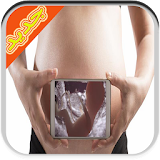


تصوير الجنين اثناء الحمل prank description
يمكنكم تطبيقنا من اخد صور للجنين ب استعمال الموجات فوق صوتية لكن تاكد ان الهاتف يدعم التقنية او جربي المحاولة عدة مرات .معلومات مهمة :
التصوير بالموجات فوق الصوتية يتيح لطبيبكم متابعة نمو وتطور طفلكم. التصوير بالموجات فوق الصوتية يمكن تنفيذه في أية مرحلة من مراحل فترة الحمل. هذا الفيديو يظهر صورة لجنين عمره 17 أسبوعا. يمكنكم أن تشاهدوا فيه نبضات القلب وكذلك عملية فتح وإغلاق الجنين لفمه.
يمكن اجراء التصوير بالشعة فوق الصوتية عند الحمل عبر البطن، وهي الطريقة المفضلة في الثلث الثاني والثالث من الحمل او عبر المهبل. في الثلث الاول من الحمل تعتبر الاشعة فوق الصوتية اداة التي تساعد على تحديد عمر الحمل، تمثيل نبض الجنين، تحديد موقع الحمل، وتحديد عدد اكياس الحمل في الحمل متعدد الاجنة. في الثلث الثاني والثالث من الحمل يتم اجراء متابعة فوق صوتية لابعاد الجنين، موقع المشيمة، ومعطيات التي تمثل وضعه الجيد، ككمية الماء وحركته. التصوير بالاشعة فوق الصوتية هو اداة ضرورية لاجراء العمليات الباضعة، بهدف التشخيص الجيني السابق للولادة وبهدف علاج الجنين في الحالات المهددة لحياته في الرحم. فحص زغب المشيمة (Chorionic Villus Sampling-CVS) يجرى في الاسابيع ال 11-12 للحمل، عن طريق المهبل او عبر البطن، بواسطة ابرة والتي يتم ادخالها لمنطقة المشيمة بهدف سحب زغب المشيمة. بمساعدة مستنبت خلايا المشيمة يمكن الحصول على معلومات متعلقة بالجينات الجنينية، وبامراض وراثية كالتليف الكيسي (Cystic Fibrosis)، هيموفيليا (Hemophilia)، متلازمة الـ (x) الهش (Fragile X Syndrome)، مرض تاي زاكس (Tay - Sachs Disease) وغيرها. يتم اجراء فحص السائل السلوي (Amniocentesis) ابتداء من الاسبوع ال 16 للحمل عبر البطن.
فحص التشوهاتات
هذا الفحص بالموجات فوق الصوتية مفصل، يتنفذ عادة في مرحلة الحمل بين 18 أسبوعا و 21 أسبوعا. يتحقق الفحص من أي تشوهات هيكلية رئيسية (فيزيائية) في الجنين، على الرغم من أنها لا تستطيع أن تظهر جميع
المشاكل.
ويتم فحص التشوهات بنفس الطريقة التي يتم بها الفحص الأول، مع مرهم على بطنك و يقوم الشخص الذي يجري الفحص بتمرير الجهاز الذي يطلق الأمواج فوق الصوتية إلى الأمام والخلف. في بعض الأحيان، فقد يكون الشخص
الذي يقوم بالتصوير هادئا حيث أنه يركز على فحص طفلك. ومع ذلك، فإنه سيكون قادرا على التحدث معك عن الصور فور الانتهاء من الفحص. معظم المستشفيات ترحب بالشركاء في غرفة الفحص. ولكن تحتاجين للتحقق من هذا مع
المستشفى الخاص.
التصوير لتحديد موعد الولادة
قد يطلب منك عدم الذهاب للحمام (التبول) قبل التصوير بالأمواج فوق الصوتية. تدفع المثانة الممتلئة رحمك للأعلى، مما يعطي صورة أفضل.
تستلقين بعد ذلك على ظهرك ويتم وضع بعض الجل الزلق على بطنك. و يتم تمرير جهاز صغير إلى الخلف وإلى الأمام على بشرتك، يبث أمواج صوتية عالية التردد خلال البطن وإلى الرحم. وينعكس الصوت ويخلق صورة تظهر على
شاشة تلفزيون.
اطلبي أن يتم شرح الصورة لك إذا بدت مربكة. وينبغي أن يكون من الممكن لشريك حياتك أن يأتي معك ويرى المسح الضوئي. كثير من الأزواج يشعرون أن هذا الفحص يساعد على جعل الطفل يبدو حقيقيا بالنسبة لهم. قد
تكونين قادرة على الحصول على صورة لجنينك- قد يكون هناك رسم مادي صغير لهذا الغرض.
ما هو مجال استخدام الاشعة فوق الصوتية؟
يمكن استخدام التصوير بالموجات فوق الصوتية في عدة طرق:
• للتحقق من حجم الجنين. يتم هذا في التصوير لتحديد موعد الولادة، يعطي هذا فكرة أفضل عن كم عدد أسابيع الحمل لديك. وسيتم تحديد موعد ولادتك وفقا لنتائج فحص الموجات فوق الصوتية.
• للتحقق ما إذا كنت تحملين أكثر من جنين واحد.
• للكشف عن معظم حالات التشوهات.
• لإظهار وضع الجنين والمشيمة. على سبيل المثال، إذا كانت المشيمة منخفضة إلى أسفل في الفترة الأخيرة من الحمل، قد ينصح بإجراء عملية قيصرية.
• للتأكد من نمو الجنين بشكل طبيعي (وهذا مهم بشكل خاص إذا كنت حاملا بتوأم أو كان لديك مشاكل في هذا الحمل أو الحمل السابق).
التصوير بالموجات فوق الصوتية يستخدم هذه الأمواج لتكوين صورة للطفل في الرحم. هذه الفحوص غير مؤلمة أبدا، وليس لها أي آثار جانبية معروفة على الأمهات أو الأجنة ويمكن القيام بها في أي مرحلة من الحمل.
تحدثي مع القابلة، أو طبيب التوليد عن أية مخاوف لديك.
يمكن لهذه الفحوص الكشف عن بعض التشوهات الهيكلية الخطيرة لذلك يجب أن تكوني مستعدة لهذه المعلومات، على الرغم من أن غالبية عمليات الفحص تبين أن الجنين ينمو بشكل طبيعي.
و تقدم كلا الصورتين لجميع الحوامل، ولكن لا تقبل جميعهن الخضوع لهما. سيتم احترام اختيارك إذا قررت عدم الحصول على الفحص، وسوف تعطى لك الفرصة لمناقشة الأمر مع فريق الأمومة الخاص بك قبل اتخاذ قرارك.
important information :
Ultrasound imaging allows your doctor to follow the growth and development of your child. Ultrasonography can be implemented at any stage of pregnancy. This video shows the picture of a fetus 17 weeks old. You can see the heart as well as the process of opening and closing the mouth of the fetus.
You can hold Balhah ultrasound imaging at conception through the abdomen, which is the preferred method in the second and third trimesters of pregnancy, or through the vagina. In the first trimester of pregnancy is the ultrasound scan tool that helps to determine gestational age, represent the pulse of the fetus, determine pregnancy site, and determine the number of bags of pregnancy in a multi-embryo pregnancy. In the second and third trimester of pregnancy is a follow-up ultrasound to keep the fetus, placenta, and data representing a good position, Kkamih water and movement. Imaging ultrasound is an essential tool for invasive operations, the aim of the previous genetic diagnosis for the birth and the aim of the fetus in the endangered his life in the womb cases treatment. Fluff examination of the placenta (Chorionic Villus Sampling-CVS) being in the 11-12 weeks of pregnancy, through the vagina or through the abdomen, and by a needle that is inserted for the placenta in order to pull the fluff placenta. With the help of cultured placental cells can be obtained information relating to embryonic genes, and disease and hereditary cystic Kaltlev (Cystic Fibrosis), Hemophilia (Hemophilia), syndrome of (x) the fragile (Fragile X Syndrome), Tai disease Sachs (Tay-Sachs Disease) and others. Is an examination of amniotic fluid (Amniocentesis) starting from the 16th week of pregnancy through the abdomen.
Examination Alichohatat
This ultrasound examination detail, usually Atnfz in pregnancy stage between 18 weeks and 21 weeks. Examination achieved any major structural defects (physical) in the fetus, although they can not
all the problems appear.
The distortions are examined in the same way in which the first examination, with the ointment on your stomach and the person conducting the examination by passing the device, called ultrasound
forward and backward. Sometimes, it may be the person doing the filming quiet as it focuses on the child's examination. However, it will be able to talk to you about the pictures immediately after
the completion of the examination. Most hospitals welcomes partners in the examination room. But you need to check this with the private hospital.
Imaging to determine the date of birth
You may be asked not to go to the bathroom (urinating) by ultrasonography. A full bladder is the highest paid of the uterus, which gives a better picture.
Tstgayn post it on your back and do some slippery gel put on your tummy. And a small device is passed back and forth on your skin, transmits sound waves of high frequency through the abdomen and into
the uterus. The reflected sound and creates an image appears on the TV screen.
Ask to be explained to you if the picture seemed confusing. It should be possible for your partner to come with you and see the scan. Many couples feel that this examination helps to make the baby
seem real for them. You may be able to get a picture of Jnenk- there may be a small material fee for this purpose.
What is the use of ultrasound?
You can use ultrasound in a number of ways:
• To check the size of the fetus. This is made in imaging to determine the date of birth, this gives a better idea of how many weeks of pregnancy you have. And will set the date of your birth,
according to the results of screening ultrasound.
• To check whether you are more than one embryo are carrying.
• to detect most cases of deformities.
• To show the fetus and placenta develop. For example, if the placenta is low down in the last period of pregnancy, it may advise a Caesarean section.
• To ensure the growth of the fetus naturally (and this is especially important if you are pregnant with twins or you have problems in this pregnancy or previous pregnancy).
Ultrasound imaging uses these waves to form a picture of the baby in the womb. This testing is not painful at all, and have no known side effects on mothers or embryos and can be performed at any
stage of pregnancy. Talk to your midwife or obstetrician for any of your concerns.
These tests can detect some serious structural defects so you have to be ready for this information, despite the fact that the majority of the checks show that the baby is developing normally.
And it offers both the two images to all pregnant women, but do not accept all of them succumb to them. We will respect your choice if you decide not to get the test, and you will be given the
opportunity to discuss it with your maternity team before making your decision.